01
Image Input
Analyze blastocyst imagery using an AI-assisted image analysis workflow.
BLASTO-VD AI analyzes blastocyst images and provides a structured grading result, bringing advanced AI-assisted image analysis into the embryology workflow.
Blastocyst • Morphology • Grading
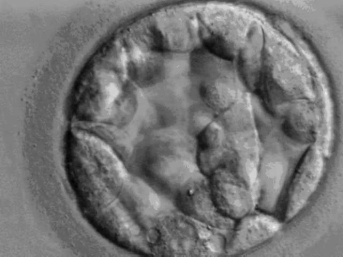
01
Image Input
02
Feature Pass
03
Grading
Interface graphics are conceptual. Microscopy photograph: Wikimedia Commons.
What is BLASTO-VD AI
BLASTO-VD AI is an artificial intelligence system designed to analyze blastocyst images and provide a grading result from image-based morphological assessment.
01
Analyze blastocyst imagery using an AI-assisted image analysis workflow.
02
Computer vision models examine visual characteristics within the provided image.
03
Receive a structured grading result to support the embryology workflow.
How It Works
Step 01
Provide a blastocyst image.
Step 02
BLASTO-VD AI processes the image using computer vision.
Step 03
The system evaluates relevant morphological features.
Step 04
A grading result is generated for review.
Blastocyst Analysis
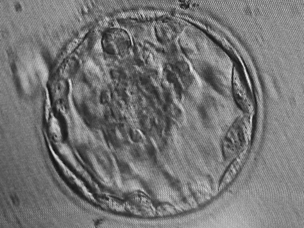 Microscopy Input
Analysis Flow
Demo / Illustrative OutputImage
Blastocyst image provided
Morphological Analysis
Visual characteristics examined
AI Processing
Computer vision evaluation
Grading Output
Structured result generated
Example result panel
Grading result
—
Status
Awaiting image
This panel is an illustrative interface concept. It does not represent a patient result and is not a clinical output.
Technology
BLASTO-VD AI applies image-based artificial intelligence to blastocyst imagery. The system reads the image, evaluates morphological features, and returns a structured grading result intended to support — not replace — the judgement of qualified embryologists.
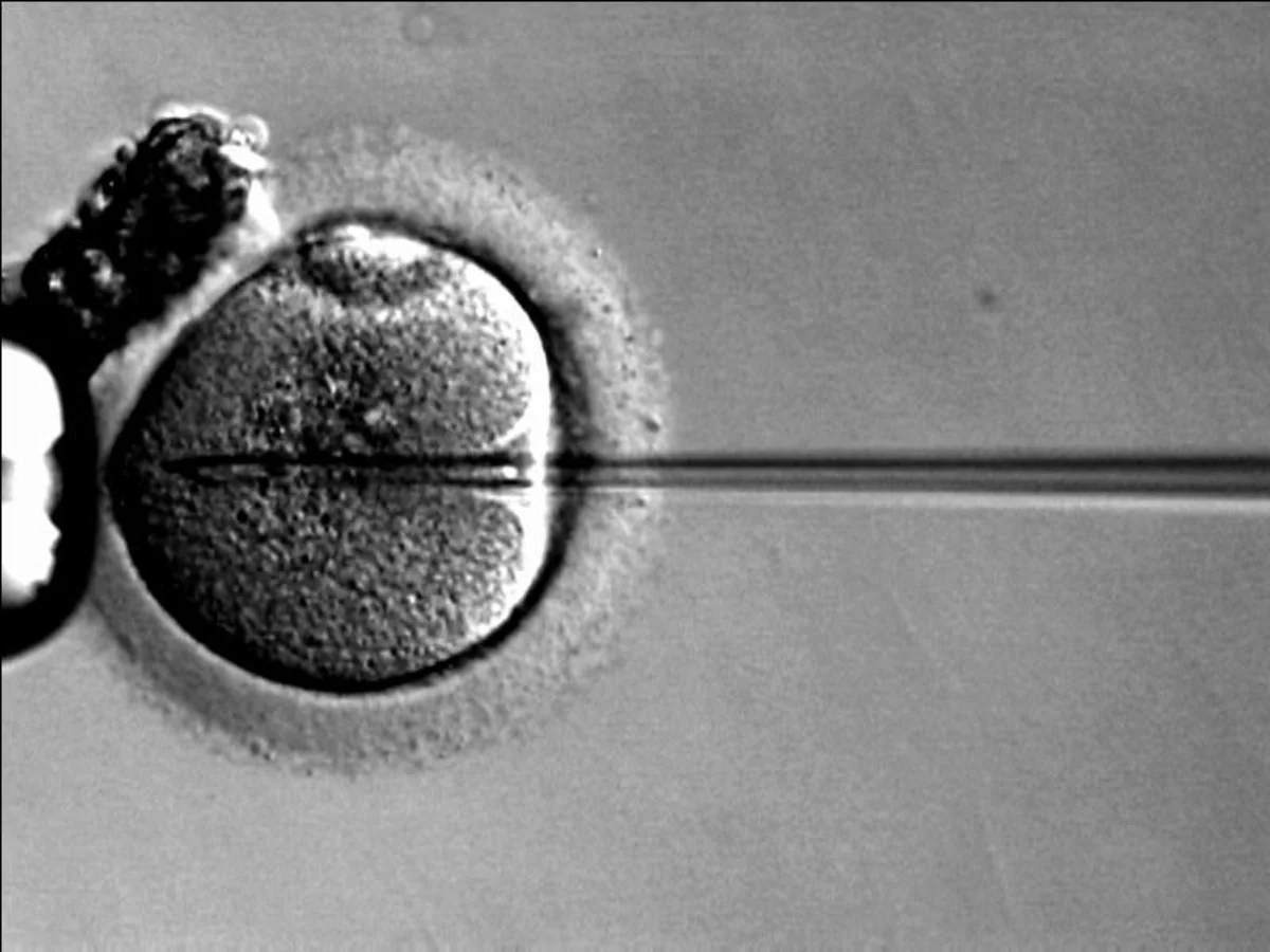 Embryology Laboratory Imaging
Image-based models trained to read visual structure.
Blastocyst imagery is processed as visual data.
Relevant morphological features are evaluated.
A grading result is produced from the analysis.
Results are returned in a consistent, reviewable form.
Why BLASTO-VD AI
Works directly from blastocyst imagery, no additional inputs required.
Returns a consistent grading result in a reviewable format.
Computer vision supports assessment within the existing workflow.
Designed around the way fertility laboratories already work.
Collaboration
BLASTO-VD AI is developed in collaboration with Prakriti Fertility Clinic and Voyed.

Prakriti Fertility Clinic
Voyed
Contact
Interested in learning more about BLASTO-VD AI or exploring access for your clinic or laboratory? Get in touch with our team.